Brain surgeons use a variety of high-tech devices and gadgets every day in the operating room, employing classic, standard tools as well as innovative ones to diagnose and treat patients. Brain surgeons, also called neurologists, are responsible for diagnosing and treating brain injuries and illnesses. They conduct neurological exams, gather medical history, do research on the latest technologies, prescribe medicine, interpret tests, and perform surgeries. The outlook for this particular job sector is good, with a higher-than-average growth of 24 percent from now until 2020, according to the Bureau of Labor Statistics.

Gadgets and Devices
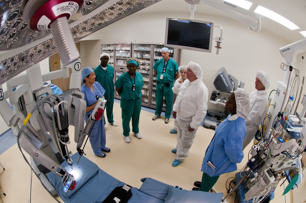
From hematoxylin and eosin (H&E) staining (designed for diagnosing brain tumors) to laser microscopes, brain surgery instruments are common in the operating and emergency rooms. Common instruments include endoscopes, MRI and CT scan equipment, scalpels, forceps, blades, retractors, hemostats, magnetic stereotaxis systems, neurosurgery imaging and neuronavigation systems, cranial sets, high-speed drills, wire saws, suctions, scalp clips, sterilizers, lab dishes, and bone punches.
Neurologists have at their disposal the latest in innovative technologies to treat and diagnose brain disease and tumors. One such fairly recent development is gamma knife radiosurgery, which is essentially a kind of radiation therapy that zaps tumors and other abnormalities such as arteriovenous malformations (AVMs). High-tech equipment shoots 200 small beams of radiation on the target in the brain, a procedure touted for its high precision and accuracy. Healthy tissues surrounding the problem area are left relatively unharmed, so the gamma knife procedure is thought to be much safer and more efficient than traditional brain surgery. It’s also pretty quick to administer; often, patients are in and out within a day.

Innovative Tools
In an effort to more aptly concentrate on areas of the brain to be operated on, a recent development in the world of neurological tools involves the use of real-time brain images and guidance that’s perfected in steps. The practice is gaining momentum and is currently being used in 21 American hospitals, according to an article in Businessweek. It essentially allows surgeons to get a better view of what they are working on. In the past, patients had to remain conscious while their doctor poked around their brains for feedback. Usually brain surgeons get an image of the brain, pinpoint the area in need of work, and cut through the skull to access it. Though not in pain, the patient would have to remain awake for the procedure, allowing for an uncomfortable experience to say the least. With this new technology, surgeons can cut operation times by about two-thirds, and the additional good news for patients is they don’t have to be conscious for the procedure. An MRI machine sends images of the brain to a monitor where brain surgeons zero in on the problem area. The innovative technology is a software that creates a map of sorts, showing doctors how to get from the skull surface to the pinpointed area. Not only that, it shows which areas to avoid (such as blood vessels) that could cause a big problem if nicked. The patient wears a helmet called a smart grid, which gives spot-on coordinates for entry. The surgeon can then use the built-in tools to navigate the brain.
Each year, more and more innovations hit the market, allowing brain surgeons the ability to further hone their craft and help heal patients in need.
Byline
Dennis Montagu writes on medicine, medical research, medical technology, the medical profession, healthcare and other related topics. Those who’d like to join the medical field should consider taking CNA Classes as a possible means of entry.
Image credit goes to Army Medicine.